Abstract: This primer explains the neurobiological effects of toxic stress in the brain and the detrimental outcomes that psychological trauma can have on the development of both mental and physical health in young adults.
"Neurobiological Underpinnings for Trauma-Informed Care: A Primer" (2015)
By Julie M. Rosenzweig
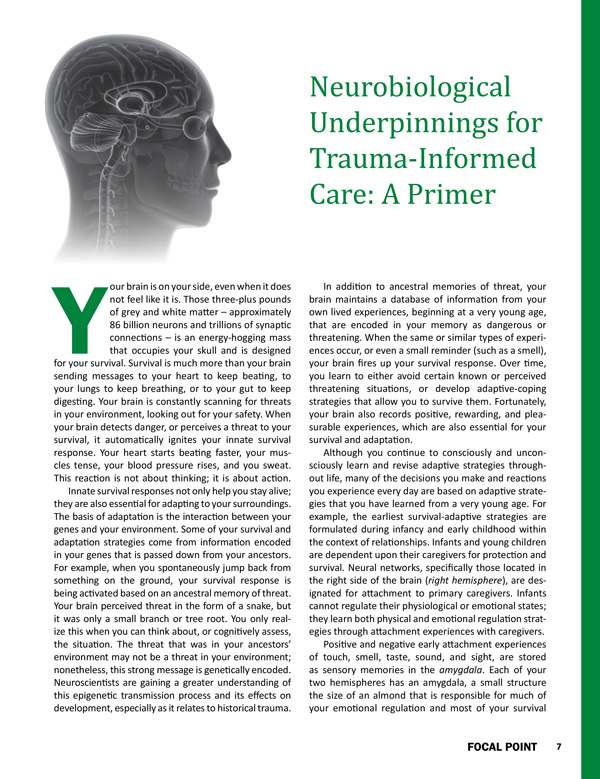
Your brain is on your side, even when it does not feel like it is. Those three-plus pounds of grey and white matter – approximately 86 billion neurons and trillions of synaptic connections – is an energy-hogging mass that occupies your skull and is designed for your survival. Survival is much more than your brain sending messages to your heart to keep beating, to your lungs to keep breathing, or to your gut to keep digesting. Your brain is constantly scanning for threats in your environment, looking out for your safety. When your brain detects danger, or perceives a threat to your survival, it automatically ignites your innate survival response. Your heart starts beating faster, your muscles tense, your blood pressure rises, and you sweat. This reaction is not about thinking; it is about action.
Innate survival responses not only help you stay alive; they are also essential for adapting to your surroundings. The basis of adaptation is the interaction between your genes and your environment. Some of your survival and adaptation strategies come from information encoded in your genes that is passed down from your ancestors. For example, when you spontaneously jump back from something on the ground, your survival response is being activated based on an ancestral memory of threat. Your brain perceived threat in the form of a snake, but it was only a small branch or tree root. You only realize this when you can think about, or cognitively assess, the situation. The threat that was in your ancestors' environment may not be a threat in your environment; nonetheless, this strong message is genetically encoded. Neuroscientists are gaining a greater understanding of this epigenetic transmission process and its effects on development, especially as it relates to historical trauma.
In addition to ancestral memories of threat, your brain maintains a database of information from your own lived experiences, beginning at a very young age, that are encoded in your memory as dangerous or threatening. When the same or similar types of experiences occur, or even a small reminder (such as a smell), your brain fires up your survival response. Over time, you learn to either avoid certain known or perceived threatening situations, or develop adaptive-coping strategies that allow you to survive them. Fortunately, your brain also records positive, rewarding, and pleasurable experiences, which are also essential for your survival and adaptation.
Although you continue to consciously and unconsciously learn and revise adaptive strategies throughout life, many of the decisions you make and reactions you experience every day are based on adaptive strategies that you have learned from a very young age. For example, the earliest survival-adaptive strategies are formulated during infancy and early childhood within the context of relationships. Infants and young children are dependent upon their caregivers for protection and survival. Neural networks, specifically those located in the right side of the brain (right hemisphere), are designated for attachment to primary caregivers. Infants cannot regulate their physiological or emotional states; they learn both physical and emotional regulation strategies through attachment experiences with caregivers.
Positive and negative early attachment experiences of touch, smell, taste, sound, and sight, are stored as sensory memories in the amygdala. Each of your two hemispheres has an amygdala, a small structure the size of an almond that is responsible for much of your emotional regulation and most of your survival esponses. The amygdala is like a smoke detector, or first responder, assessing incoming sensory experiences for threat. The implicit memory system is the only type of memory system that is online at birth and, in addition to encoding early life attachment experiences, is responsible for encoding traumatic experiences across the lifespan. Around 18 months of age, the explicit memory system, a function of a brain structure called the hippocampus, begins to develop. The hippocampus is particularly good at remembering place (spatial) and time details about everyday activities. The amygdala and the hippocampus are located next to one another and communicate consistently. When the amygdala registers an emotional experience, the hippocampus provides details of the effect.
You are most familiar with your hippocampal-based explicit memory system, as you use it to consciously recall information; hence it is crucial to learning. The hippocampus works closely with the prefrontal cortex (PFC) through neural pathways to create long-term memory storage. Best known for its role in executive functioning, including decision-making, inhibition, and anticipating consequences, the PFC, located in the exterior part of the front of the brain just below the skull and behind the forehead, is also active in affective regulation. Interestingly, the PFC is the last area of the brain to become fully functional, typically in the mid to late 20s.
Information encoded in the implicit memory system is not remembered in the same way. An implicit memory, also called an unconscious memory, is based on past experiences, surfaces without thinking, and is activated by a sensation, such as a smell or a sound, and often without your conscious awareness. When an implicit memory is present, you often have an emotional response, even if you do not understand why.
Your early attachment experiences create neural patterns: specific pathways which guide your thoughts about your self-worth, how others will respond to you, and the level of safety you feel about your environments. This is your first adaptive-survival strategy and it becomes generalized to many situations, especially social and romantic relationships. In many ways, this adaptive strategy is similar to having your own signature GPS (global positioning system), that helps you navigate relationships and real or perceived threats in your environment. Because humans are social animals, your survival and well-being depends on feeling safe with others. The most powerful resources in stressful or threatening situations are other people. Early attachment relationships that provide you with experiences of safety, protection, and caring equip you to trust others to do the same. If early attachment relationships are less optimal, you will be more hesitant to trust, both yourself and others.
Toxic Stress: When Adaptive-Survival Strategies Get Stuck
Knowledge about the neurobiology of threat assessment, adaptation, and attachment is the foundation for understanding how consistently experiencing adversity (chronic stress), including psychological traumatic event exposure, changes areas of the brain both structurally and functionally. The ability of the brain to change and reorganize in response to experiences is known as neuroplasticity. Neuroplasticity is essential to our survival and adaptation. It is also essential to our resilience and healing from the effects of toxic stress.
In response to all types of stress (e.g., positive, physiological, temporary, chronic, or traumatic), the Hypothalamic-Pituitary-Adrenal (HPA) axis becomes activated. When your HPA system is engaged, the brain prioritizes resources for activities that are essential for survival, such as heart rate, blood pressure, and muscle tension, while processes such as digestion and higher cognitive functions not involved in fight or flight are minimized. Once the amygdala sends the message that a threat is imminent or present, the Hypothalamus releases corticotropin-releasing hormone (CRH), which signals the Pituitary gland to release adrenocorticotropic hormone (ACTH), which prompts the Adrenal gland to release cortisol. The HPA axis is the body's stress management system and coordinates not only with the amygdala, but also with the hippocampus and the prefrontal cortex (PFC). While the amygdala sounds the alarm, the hippocampus and the PFC shut the alarm off.
When the HPA axis is continually activated by threat or chronic stressors in a person's environment, the brain adapts to these environmental demands by remaining on high alert, ready for action at all times. The body cannot effectively metabolize the high levels of cortisol (toxic stress), and this heightened, persistent level of stress response becomes toxic to the system. Deficits or alterations in the stress regulatory processes are the basis of many mental health impairments, including post-traumatic stress and complex trauma disorders.
Toxic stress profoundly impairs the HPA system and alters the structure and functioning of brain areas. The hippocampus appears exceptionally vulnerable to the effects of stress. When there is chronic over-activation of the stress response system resulting in the brain being flooded with excess cortisol, hippocampal-dependent learning and memory processes become impaired. When altered by toxic stress, the hippocampus has a significantly reduced capacity to provide important contextual information to the amygdala about which conditions represent danger and which represent safety. When the hippocampus is compromised, it is not effective in sending messages to the hypothalamus to stop producing CRH and turn off the stress response.
Likewise, the amygdala is also changed structurally and functionally by toxic stress. The amygdala is central to relational processes and development of attachment strategies, making it extremely sensitive to excessive activation in early life. Infants, children, and youth are particularly sensitive to toxic stress and impairment in regulatory functioning. Again, excessive activation in the areas that respond to emotions, and under-activation in areas involved in cognitive processes of assessment and evaluation, lead to developmental disruptions.
Functionally, when the brain detects that the threats or stressors, or the perception thereof, have been stopped or prevented, the stress system response is shut off, deactivating the cascade of chemicals and returning the brain and HPA axis to baseline or what is called homeostasis. However, when the threat is frequent or prolonged, the ability of the brain and HPA axis to return to homeostasis becomes impaired.
Because of this activated state, sensory information associated with the threat or chronic stressors, such as visual cues, smells, ambient sounds, or the time of day, continue to stay active in the amygdala. As a result, negative affective states (e.g., fear, shame, rage, numbing, dissociation) and physical sensations (e.g., muscle tension, increased heart rate, rapid breathing) paired with the sensory cues are easily activated even in seemingly non-threatening environments.
For example, a high school student is jumpy, feels her heart beating, and experiences the emotion of fear at the sound of a loud noise in the hallway, even though she is in a safe classroom environment with a teacher and friends she trusts. Her amygdala holds implicit memories that include loud sounds, such as gunshots or parents yelling at each other. The loud sound in the hallway represents threat and the amygdala sends a message to the HPA to release more chemicals. The hippocampus (sometimes in partnership with the PFC), may be able to intervene and slow the stress response. Based on explicit memories, the hippocampus is able to very quickly sort through its database and send a message to the amygdala that the noise is the sound of a student's locker closing and shuts off the stress response. If however, the student has chronic stressors, ongoing trauma, or complex developmental trauma, the hippocampal-based regulatory processes are significantly compromised and the student may already be enacting the survival response. For youth who are frequently in survival mode, acting on fight or flight or freeze responses, this survival-based adaptive functioning becomes a normative way of being in the world. Their brains have learned ways of protection from actual or perceived threat. In a very real way, the brain has become trauma-informed.
Trauma-informed care, whether provided by individuals or organizations, recognizes the pervasiveness of adverse experiences among service users and the real effects of toxic stress on their mental and physical health. Although these effects can be damaging, through understanding the brain's neuroplasticity and its biological imperative for relational connection, there is an abundance of hope for healing and transformation.1
References
- McEwen, B. S., Gray, J. D., & Nasca, C. (2015). Recognizing resilience: Learning from the effects of stress on the brain. Neurobiology of Stress, 1, 1-11. Retrieved from http://www.sciencedirect.com/science/article/pii/S2352289514000022
Suggested Citation
Rosenzweig, J. M. (2015). Neurobiological Underpinnings for Trauma-Informed Care: A Primer. Focal Point: Youth, Young Adults, and Mental Health, 29, 7-9. Portland, OR: Research and Training Center for Pathways to Positive Futures, Portland State University.